Contact
 Hoe winkelen bij ons werkt
Bezorging
Winkelgids
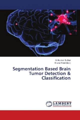
Segmentation Based Brain Tumor Detection & Classification
 Engels
 86 b
86 b
Retourneren binnen 30 dagen
Klanten kochten ook


 /
/
 Gebonden (paperback)
 24.17
€
24.17
€


Biomedical Image Processing consists many different types of imaging methods likes CT scans, X-Ray and MRI. These techniques allow humans to identify even the smallest abnormalities in the human body. The primary goal of medical imaging is to extract meaningful and accurate information from the images with the least error possible. MRI (Magnetic Resonance Imaging) is a medical technique, mainly used by the radiologist for visualization of internal structure of the human body. MRI provides useful information about the human soft tissue, which helps in the diagnosis of brain tumor. Image segmentation refers to partitioning of image into multiple regions or segments such that it can meaningfully represent the image through which information can be extracted. In this paper we are using Canny and SIFT techniques for segmentation of brain image considering shape and texture features. After that Support Vector Machine (SVM) is used to classify tumor and non-tumor regions. The performance of the proposed method is evaluated in terms of Sensitivity (Se), specificity (Sp), precision (Pr) and accuracy (Acc) and PSNR.


Informatie over het boek
Engels
Categorieën

Geef dit boek vandaag nog cadeau
Dat gaat heel eenvoudig
1 Voeg het boek toe aan je winkelwagentje en selecteer Als cadeau bezorgen 2 Je krijgt van ons per omgaand een voucher 3 Het boek wordt bezorgd op het adres van de ontvangerDit vind je misschien ook interessant

 /
Gebonden (paperback)
16.48
€
/
Gebonden (paperback)
16.48
€